The Headache That Hid a Brain Tumor for Over a Decade
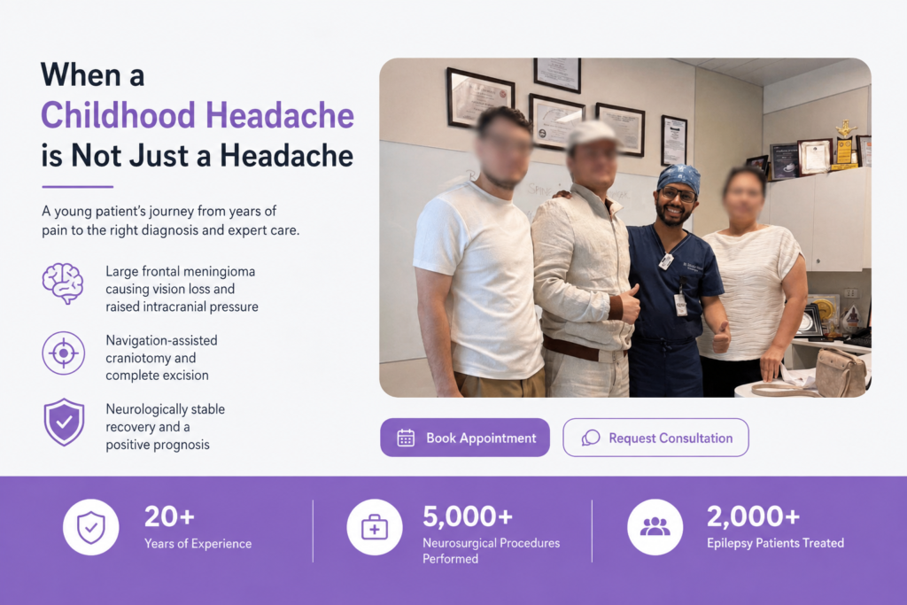
Mr. Sulaymon Ibrokhimov had been living with occipital and bifrontal headaches since 2012. Five months before reaching Dr. Satyakam Baruah in Faridabad, he developed transient blackouts of vision in both eyes, which turned into painless persistent vision loss one week before admission. Imaging revealed a large left frontal convexity meningioma with mass effect. He underwent a navigation-assisted left frontal craniotomy and excision of the tumor by Dr. Satyakam Baruah on 27 February 2026 and was discharged neurologically stable on 4 March 2026.
Early Diagnosis Saves Lives - Book an Appointment Today with Dr Satyakam Baruah!
When a childhood headache is not just a headache
Mr. Sulaymon’s headaches were not new. They had begun in 2012, when he was about nine years old. The pain was dull and aching, predominantly localised to the occipital and bifrontal regions, the back of the head and across the forehead. It was persistent, with intermittent flare-ups.
Like most children, he adjusted. Over-the-counter painkillers offered temporary relief. At one point he was evaluated in Uzbekistan, where the MRI was verbally reported to be normal, and he was treated for idiopathic intracranial hypertension, a condition where pressure inside the skull is raised without a clear structural cause. With that label in place, the headaches were accepted as part of his life.
For more than a decade, no one suspected that something was slowly growing inside his skull.
What are the warning signs of raised intracranial pressure?
In the six months before he came to Dr. Satyakam Baruah, the pattern shifted. The headache became worse in the early morning and was aggravated by straining, bending forward, or sudden changes in posture. The analgesics that had always offered some relief began to fail.
These are recognised in neurosurgery as classic signs of raised intracranial pressure. The skull is a closed box. If something inside it begins to take up space, the pressure rises, and the body shows it in specific ways. Morning headaches and posture-related pain are two of the clearest signals. His headache also had none of the features of migraine, no photophobia, no throbbing quality, no aura, and no significant nausea or vomiting.
Why vision changes should never be ignored
About five months before admission, Mr. Sulaymon began experiencing transient episodes of visual loss in both eyes simultaneously. Each was sudden in onset, lasted a few seconds, and resolved on its own. Easy to explain away as tiredness. Then, one week before he reached the hospital, he developed acute, painless loss of vision in both eyes that did not resolve. Painless vision loss does not feel like an emergency, but it almost always is.
On evaluation, he was found to have papilledema, a swelling of the optic discs that confirms raised pressure inside the skull. Following ophthalmic and pre-anaesthesia assessments, imaging revealed the underlying cause: a large left frontal convexity meningioma with mass effect, causing the papilledema-related changes affecting his vision.
What is a meningioma and why is it dangerous?
A meningioma is a tumor that grows from the meninges, the protective coverings of the brain and spinal cord. Most are benign and slow-growing, which is both their strength and their danger. Because they grow gradually, the brain adapts and symptoms can stay vague for years. By the time they become obvious, the tumor is often large.
In Mr. Sulaymon’s case, the Brain tumor was a large, extra-axial, highly vascular mass arising from the anterior frontal convexity on the left side. It was compressing the underlying brain, had infiltrated the pia (the delicate layer covering the brain surface), and had pushed intracranial pressure to the point where his optic nerves were beginning to suffer. Without surgery, permanent blindness was likely.
Navigation-assisted brain tumor surgery in Faridabad
On 27 February 2026, Consultant Neurosurgeon Dr. Satyakam Baruah and his team performed a navigation-assisted left frontal craniotomy and excision of the meningioma under general anaesthesia. Navigation-assisted surgery works like a GPS for the brain. Frameless neuronavigation uses pre-operative imaging to build a three-dimensional map of the patient’s anatomy that guides every step in real time. This precision mattered because the tumor had breached the dura adjacent to the superior sagittal sinus, a major venous channel where any injury can be life-threatening.
Intraoperative findings matched the imaging: a large, extra-axial, highly vascular tumor arising from the anterior frontal convexity, with pial infiltration and compression of the underlying brain. It was dural-based but not invading the sinus, and the overlying skull bone showed hyperostotic changes, a finding that often accompanies long-standing meningiomas. Dr. Baruah performed internal decompression followed by meticulous circumferential dissection from the surrounding brain. Gross total excision was achieved with full preservation of the superior sagittal sinus. Duroplasty was carried out using the patient’s own pericranial tissue, and the hyperostotic inner table of the bone flap was drilled away before the bone was replaced with plates and screws. A post-operative MRI confirmed no residual tumor.
Recovery after meningioma surgery
In the immediate post-operative period, Mr. Sulaymon was electively monitored in the Neuro ICU. On the first post-operative day, he was shifted to the ward, hemodynamically stable and neurologically preserved, with no new deficits. That same night, he developed significant post-operative headache and was shifted back to the ICU for closer monitoring and optimised pain management. He remained neurologically stable throughout, with preserved higher mental functions and no worsening of vision. Steroids were gradually tapered, and his recovery continued steadily.
By the third post-operative day, the surgical drain was removed, the operative wound was healthy with no signs of infection or cerebrospinal fluid leak, and he was being progressively mobilised. On 4 March 2026, he was discharged: conscious, alert, oriented, and ambulant with support (necessary because of his poor vision), with no new deficits. His discharge prognosis was recorded as good.
Vision recovery is a slower journey. Optic nerves that have been compressed for years take time to heal, and in some cases recovery is incomplete. But the cause has been fully addressed, and that is the foundation on which the rest of his recovery will be built.
Struggling with Seizures, Persistent Headaches, or Other Neurological Symptoms?
When should you see a neurosurgeon for a headache?
A few lessons come out of this case clearly. Long-standing headaches deserve attention, especially when their pattern changes. Morning headaches, posture-related pain, and reduced response to medication are red flags that should never be ignored. Visual symptoms in both eyes, even brief ones, are significant, often the first sign that pressure inside the skull is affecting the optic nerves. And a normal MRI in the past does not rule out a future problem. Symptoms evolve, and so do imaging findings. Fresh symptoms deserve fresh evaluation.
Consult a brain tumor specialist in Faridabad
Mr. Sulaymon walked into the hospital with failing vision and walked out with his tumor removed and a future ahead of him. If you or someone in your family has been living with long-standing headaches, vision changes, or other unexplained neurological symptoms, an early specialist opinion can make all the difference. Dr. Satyakam Baruah, Consultant Neurosurgeon in Faridabad, offers expert evaluation and treatment for brain tumors, meningiomas, and other complex neurosurgical conditions for patients across Delhi NCR.