MRI Negative Epilepsy Treatment in Faridabad
MRI Negative Epilepsy Treatment in Faridabad by Dr. Satyakam Baruah offers advanced evaluation with Video EEG, PET, SPECT, SEEG, and epilepsy-focused imaging to identify seizure sources and guide personalized treatment for patients across Faridabad and Delhi NCR.
Early Diagnosis Saves Lives - Book an Appointment Today with Dr Satyakam Baruah!
What is MRI Negative Epilepsy?
MRI negative epilepsy describes a situation where a patient has recurrent seizures, often meeting criteria for drug resistant epilepsy, but standard MRI imaging does not reveal an obvious structural abnormality. This can be confusing and frustrating for patients, since seizures clearly originate from somewhere in the brain even when imaging looks unremarkable.
There are several reasons MRI may appear normal despite ongoing seizures. Some abnormalities, such as subtle areas of cortical dysplasia, are simply too small or too similar in appearance to normal brain tissue to be identified on standard imaging protocols. Others may be present but require a higher resolution scan, a specific epilepsy protocol, or expert review by a radiologist experienced in epilepsy imaging to detect. In some cases, the seizure generating area genuinely has no visible structural correlate on any current imaging technology.
The difference between MRI negative and MRI positive epilepsy lies primarily in how the seizure focus is identified and confirmed. In MRI positive cases, a visible lesion such as mesial temporal sclerosis or a small tumor often guides diagnosis and treatment planning directly. In MRI negative cases, the team must rely more heavily on a combination of clinical history, EEG findings, and advanced functional imaging to build a picture of where seizures originate.
This is precisely why specialised evaluation matters so much for MRI negative epilepsy. A normal MRI read by a general radiologist is not the same as a comprehensive epilepsy workup performed by a team experienced specifically in localizing seizure foci that do not show up on routine scans.
Symptoms of MRI Negative Epilepsy
The symptoms of MRI negative epilepsy are not different in nature from epilepsy with visible MRI findings. What sets this group apart is the imaging result, not the seizure pattern itself.
Patients may experience focal seizures, which begin in one area of the brain and may or may not spread further. Focal impaired awareness seizures involve some alteration or loss of awareness, often with repetitive movements or behaviors the patient does not recall afterward. Some patients experience seizures that become generalised, spreading to involve both sides of the brain and sometimes leading to convulsions.
Auras, brief sensory, emotional, or cognitive experiences occurring just before a seizure, are common and can be an important diagnostic clue, since the nature of the aura often points toward the area of the brain where the seizure begins. Memory disturbances around the time of seizures, and speech changes such as temporary difficulty finding words, can also occur depending on which brain region is involved.
Loss of awareness, ranging from brief lapses to more prolonged confusion, is a hallmark of many focal seizures. Muscle jerks, whether localized or more widespread, may also be part of the presentation. Because these symptoms vary so much between individuals, a detailed description of what happens before, during, and after a seizure is one of the most valuable pieces of information a patient or family member can bring to an evaluation.
Causes and Risk Factors of MRI Negative Epilepsy
Several underlying causes can lead to MRI negative epilepsy, even though they may not be visible on standard imaging.
- Microscopic brain abnormalities are changes at a cellular level too small to be resolved by current MRI technology, even with high resolution protocols.
- Cortical dysplasia, particularly subtle or focal forms, is one of the most common causes of MRI negative epilepsy. This developmental abnormality in how brain cells are organized can be missed even on epilepsy protocol MRI when mild or focal.
- Genetic factors can contribute to epilepsy without producing any structural change visible on imaging, since the underlying issue may relate to how brain cells communicate rather than their physical structure.
- Previous brain infection, such as encephalitis, can sometimes leave subtle changes not clearly visible on standard imaging but still create a focus for seizures.
- Head injury, even without an obvious lesion on MRI, can in some cases contribute to epilepsy through changes not easily detected by routine scans.
- Unknown causes account for a meaningful proportion of MRI negative epilepsy cases, where despite thorough evaluation no definitive structural or genetic cause is identified, though this does not mean treatment options are unavailable.
Routine MRI is simply not sensitive enough to detect every possible cause of seizures, which is why advanced evaluation becomes necessary.
How is MRI Negative Epilepsy Diagnosed?
Diagnosing MRI negative epilepsy requires a structured, multidisciplinary approach that goes well beyond a single scan or consultation.
- Clinical history is the starting point, with detailed documentation of seizure semiology, frequency, triggers, and patterns noticed by the patient or family. This often provides the first clues about which part of the brain may be involved.
- Neurological examination assesses overall neurological function and helps identify subtle signs pointing toward a specific area of brain involvement.
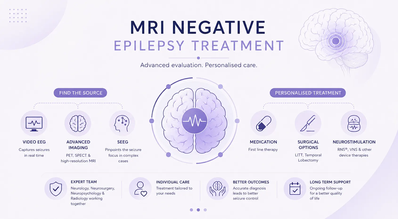
- Video EEG monitoring records brain electrical activity alongside video of the patient, correlating clinical seizure behavior with electrical activity. This is one of the most important tools in MRI negative cases, since it can help localize the seizure focus even when imaging has not.
- High-resolution MRI with epilepsy protocol uses specific sequences and slice thickness designed to detect subtle abnormalities a standard MRI might miss, often repeated with expert epilepsy-focused review even if a prior MRI was reported normal.
- PET scan can identify areas of reduced brain metabolism between seizures, which may correspond to the seizure focus even when no structural abnormality is visible on MRI.
- SPECT scan, when indicated, helps localize the seizure focus by detecting blood flow changes during or shortly after a seizure, adding another layer of information in complex cases.
- MEG, where appropriate and available, maps the magnetic fields generated by brain activity and can add further precision to localization in complex epilepsy centers.
- Neuropsychological assessment evaluates memory, language, and other cognitive functions, supporting localization and establishing a baseline relevant to treatment planning.
- Stereo EEG (SEEG) in selected patients involves placing electrodes directly within the brain to record activity from deep and widespread areas simultaneously. This invasive but highly informative technique is considered when non-invasive testing has narrowed the search but not definitively localized the seizure focus, and plays a central role in surgical planning for many MRI negative cases.
The importance of a multidisciplinary epilepsy team cannot be overstated here, since diagnosing MRI negative epilepsy depends on integrating findings from several tests, none conclusive alone, through joint review by neurology, neurosurgery, neuropsychology, and neuroradiology.
Treatment Options for MRI Negative Epilepsy
Treatment for MRI negative epilepsy is individualised based on the findings of the evaluation described above, and is not limited to surgery alone.
- Anti-seizure medications remain the first line of treatment for most patients, and many people with MRI negative epilepsy achieve good seizure control with appropriately chosen and dosed medication.
- Lifestyle and seizure management strategies, including sleep regulation, trigger avoidance, and stress management, support medical treatment for many patients.
- Epilepsy surgery evaluation becomes relevant when seizures continue despite adequate medication trials. Even without a visible MRI lesion, advanced evaluation including video EEG, PET, SPECT, and sometimes stereo EEG can identify patients who may still benefit from surgery.
- Laser Interstitial Thermal Therapy (LITT), in selected patients where the seizure focus can be confidently localized through advanced evaluation, offers a minimally invasive option for treating the identified area. Learn more on our LITT procedure page.
- Temporal lobectomy, when appropriate, remains an option for some MRI negative patients whose evaluation localizes the seizure focus to the temporal lobe despite the absence of a visible lesion.
- Responsive neurostimulation, if available, is a device-based option that can detect abnormal electrical activity and deliver targeted stimulation to interrupt seizures, sometimes considered when resection is not feasible.
- Vagus nerve stimulation (VNS) is another device-based option that can reduce seizure frequency in some patients, particularly when resective surgery is not appropriate or as an additional therapy alongside medication.
Treatment is never one-size-fits-all in MRI negative epilepsy. The path forward depends entirely on what comprehensive evaluation reveals about each patient’s seizure pattern and likely focus.
When is Surgery Considered for MRI Negative Epilepsy?
Surgery remains a meaningful option for some patients with MRI negative epilepsy, though the path to that decision is more involved than in cases with a clearly visible lesion.
Surgery is generally considered once epilepsy is classified as drug-resistant, meaning seizures have not responded to two or more appropriately chosen medications. Seizure localisation is the central challenge in MRI negative cases, requiring the combination of video EEG, advanced imaging, and sometimes stereo EEG described earlier to build enough confidence about where seizures begin.
Presurgical evaluation in these cases is typically more extensive than in MRI positive epilepsy, since the team cannot rely on a single visible lesion to guide planning. The benefits and limitations of surgery must be weighed carefully, since outcomes in MRI negative epilepsy surgery, while meaningful for well-selected patients, are generally less predictable than in cases with a clear structural lesion.
Patient selection is therefore especially important, and only patients with strong, consistent localization across multiple tests are typically considered good surgical candidates. Shared decision-making between the patient, family, and the multidisciplinary team is central to this process, ensuring that patients understand both the potential benefits and the inherent uncertainties before proceeding with any surgical option.
Struggling with Seizures, Persistent Headaches, or Other Neurological Symptoms?
Living with MRI Negative Epilepsy
Managing day to day life with MRI negative epilepsy involves many of the same considerations as epilepsy in general, alongside the added complexity of an evaluation process that may take time.
Medication adherence is essential, since missed doses are a common trigger for breakthrough seizures even in patients on an otherwise effective regimen. Trigger management, including awareness of personal seizure triggers such as sleep deprivation, stress, or specific situations, can help reduce seizure frequency alongside medical treatment.
Sleep hygiene deserves particular attention, since poor sleep is one of the most consistent seizure triggers across many forms of epilepsy. Driving regulations vary by jurisdiction and depend on seizure-free intervals defined by local law, and should always be confirmed directly with your treating physician rather than assumed.
Employment considerations may include discussing reasonable accommodations with employers where needed, and many patients with well-controlled epilepsy continue working without significant disruption. Regular follow-up with the epilepsy care team remains important throughout, both to monitor seizure control and to revisit evaluation options if circumstances change. Mental health support is also a meaningful part of comprehensive epilepsy care, since living with unpredictable seizures can affect mood and wellbeing, and addressing this openly with the care team is encouraged.
Why Choose Dr. Satyakam Baruah for MRI Negative Epilepsy Treatment in Faridabad?
MRI negative epilepsy requires a level of diagnostic expertise that goes beyond standard epilepsy care. Dr. Satyakam Baruah is a fellowship-trained epilepsy neurosurgeon with specific expertise in complex epilepsy cases, including those where standard imaging has not identified a clear cause.
Patients undergo a comprehensive epilepsy evaluation that draws on video EEG, advanced imaging, and where needed, stereo EEG, to build the most complete possible picture of each individual’s seizure focus. Where surgery is being considered, advanced surgical planning ensures that any procedure is grounded in thorough localization rather than assumption.
Care is delivered through multidisciplinary collaboration between neurology, neurosurgery, and neuropsychology, with individualised treatment planning at every stage. Long-term follow-up continues well beyond the initial evaluation, reflecting the reality that MRI negative epilepsy often requires ongoing, patient-specific management. This comprehensive approach is available to patients at Amrita Hospital, Faridabad, serving Faridabad and the wider Delhi NCR region
Frequently Asked Questions
What is MRI negative epilepsy? expand_more
MRI negative epilepsy describes recurrent seizures occurring despite a brain MRI that appears normal on standard imaging, often because the underlying abnormality is too subtle or microscopic to be detected on routine scans.
Can I have epilepsy even if my MRI is normal? expand_more
Yes. A normal MRI does not rule out epilepsy or a treatable underlying cause. Many patients with confirmed epilepsy have MRI scans that appear unremarkable.
Why is my MRI normal despite recurrent seizures? expand_more
This can happen when the seizure generating abnormality is too small or subtle to be visible with current imaging technology, even with a specific epilepsy protocol.
How is MRI negative epilepsy diagnosed? expand_more
Diagnosis relies on detailed clinical history, video EEG monitoring, high-resolution epilepsy protocol MRI, and in selected cases PET, SPECT, MEG, or stereo EEG, reviewed by a multidisciplinary epilepsy team.
Can MRI negative epilepsy be treated? expand_more
Yes. Treatment options range from anti-seizure medications and lifestyle management to surgical options, device-based therapies like VNS or responsive neurostimulation, depending on what the evaluation reveals about each individual case.
Can seizures become controlled with medication? expand_more
Many patients with MRI negative epilepsy achieve good seizure control with appropriately selected medication. When this is not the case, further evaluation for other treatment options, including surgery, may be considered.
Is MRI negative epilepsy lifelong? expand_more
The course of epilepsy varies by individual and underlying cause. Some patients achieve long-term seizure control with medication or other treatment, while others may require ongoing management, which is best discussed based on your specific evaluation.

Dr. Satyakam Baruah’s Medical Content Team
Dr. Satyakam Baruah’s medical content team specialises in creating accurate, clear, and patient-focused healthcare content. With strong clinical understanding and expertise in technical writing and SEO, the team translates complex medical information into reliable, accessible resources that support informed decisions and uphold Dr. Satyakam's commitment to quality care.
This content is reviewed by Dr. Satyakam Baruah
Related Conditions

Epilepsy Treatment in Faridabad
Epilepsy is a manageable neurological condition. Whether you’re newly diagnosed or living with drug-resistant seizures that haven’t responded to multiple…
Epileptic Spasms Treatment in Faridabad
Epileptic spasms are brief, clustered axial jerks lasting one to two seconds, most common in infants shortly after waking, and…
Related Blogs
Is Epilepsy Hereditary?
In Summary – What You Need to Know Epilepsy is not always hereditary. About 30–40% of cases have…
Read Article →
When Seizures Don’t Stop: Drug Resistant Epilepsy
Drug resistant epilepsy is diagnosed when seizures continue despite trying at least two appropriate anti-seizure…
Read Article → WhatsApp